Spotting cancer's greasy fingerprints
26 January, 2020
Lipid droplets normally involved in storing fats have recently been revealed as key players in many cancer cell processes, including their ability to resist treatment. These lipids can now be quantitatively identified more easily than ever using a microscopy technique developed at KAUST.
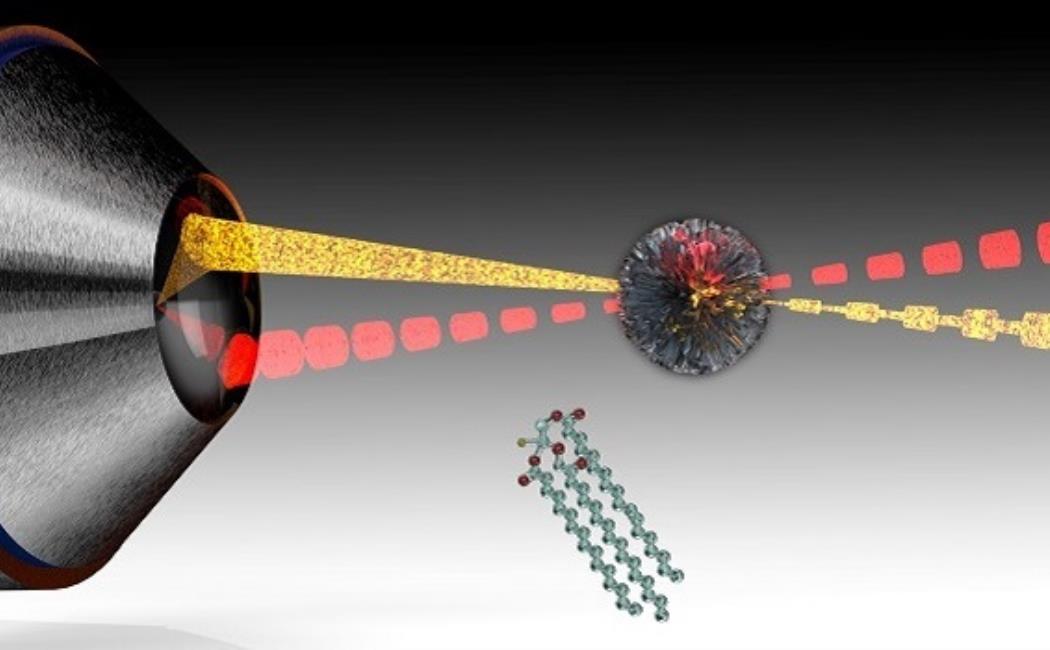
The chemical bonds holding molecules together produce unique vibrations when excited by a light source. Researchers are now exploiting this effect to characterize living cells and tissue through stimulated Raman scattering microscopy. In this approach, ultrafast laser pulses are used to vibrate biomolecules throughout the volume of a sample. The resulting signals can then help visualize components, including lipids at speeds suitable for video imaging.
Click here to read the full story
Image: Stimulated Raman microscopy of lipid droplets. Two laser beams with different colors nonlinearly interact inside the lipid droplet. The variation in their intensity holds chemical information.
© 2019 Andrea Bertoncini