The dynamic molecular changes of homing
05 August, 2018
A new microscopic technique allows researchers to observe the initial nanoscale changes that are part of the complex process of transplanted stem cells homing in on, and specifically settling in, the bone marrow. Understanding the details of this process can improve the success of stem-cell transplantations for treating leukemia.
KAUST microscopist, Satoshi Habuchi, and colleagues developed their microfluidics-based super-resolution microscopy platform to improve the ability of researchers to visualize the dynamic nanoscale changes that happen to stem cells as they move in blood. Current techniques only enable researchers to identify the molecules involved in stem cell homing—a phenomenon that allows cells to migrate to their organ of origin—but not what is happening at the nanoscale or over time.
Click here to read the full story
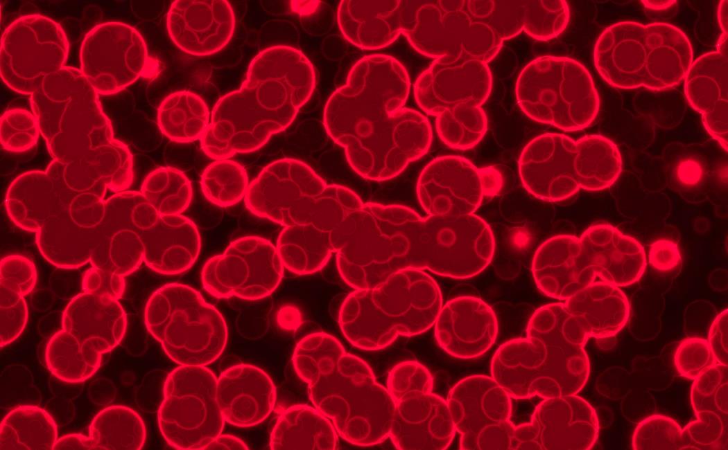
Image: HSPC homing occurs through complicated interactions between HSPCs and endothelial cells in the blood stream, which begins with selectin-ligand interactions.
© 2018 Sergiy Zhukovskyy / Alamy Stock Photo