DNA replication under the microscope
21 November, 2021
Cryogenic electron microscopy (cryo-EM) has enabled researchers to study how the DNA replication machinery assembles at sites where DNA is damaged.
Cellular DNA is continuously exposed to both endogenous and exogenous DNA-damaging agents, such as reactive oxygen species and UV radiation. To reduce the biological consequences of DNA damage, all living organisms have evolved mechanisms to tolerate and repair DNA damage to try to ensure that genetic information is accurately inherited. One such mechanism, called translesion synthesis (TLS), allows DNA replication to proceed through unrepaired DNA lesions.
TLS involves highly accurate DNA synthesizing enzymes (replicative DNA polymerases) being temporarily replaced with specialized, low-fidelity TLS polymerases that can ensure cell survival at the expense of introducing mutations. The mutagenic and translesion synthetic activity of TLS polymerases can result in normal cells becoming cancerous or cancer cells becoming drug resistant.
Click here to read the full story.
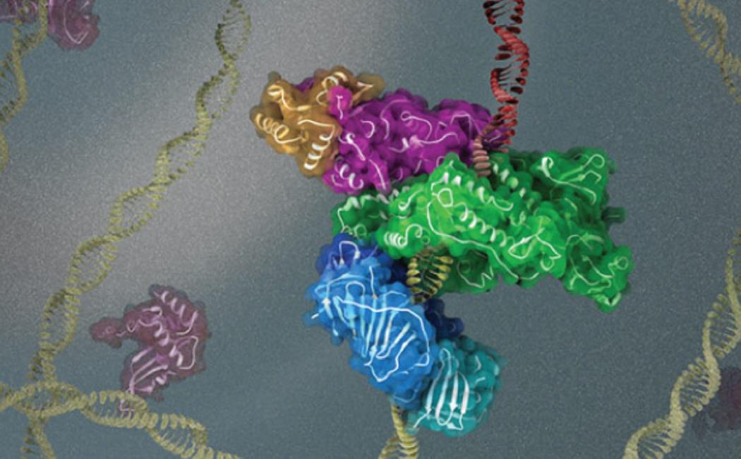
Image: KAUST scientists use cryogenic electron microscopy to investigate the 3D structure and function of key protein complexes involved in DNA replication and repair.
© 2021 KAUST; Heno Hwang